Anatomy & Physiology
Water-vascular system
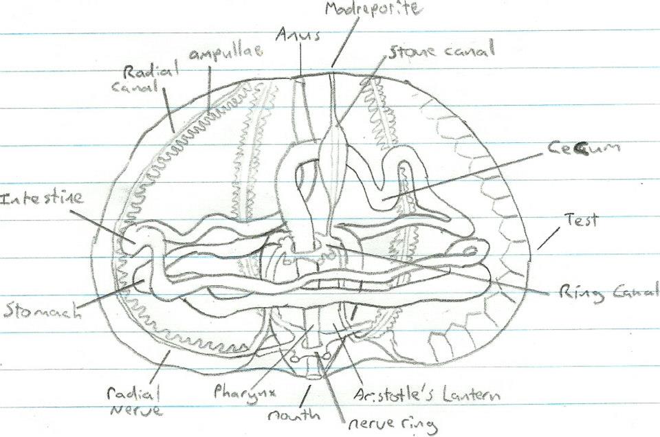
All echinoderms have a water-vascular system. This is a hemal system consisting of coelomic canals, arranged in a pentamourous configuration. The central canal, known as the ring canal, surrounds the stomach, with several radial canals branching off and running up the inside of the test along the ambulacral areas (Ruppert et al, 2004). At the end of these radial canals are small muscular sacks called ampullae, from which tube feet extend. Branching off the central ring canal, is another canal running to the aboral surface where the madreporite is found. Through the use of the madreporite and the tube feet, circulation of water and coelomic fluid can occur.

Figure 1. Anatomy of a sea urchin. Adapted from Ruppert et al, 2004.
Stomach
From the pharynx, the oesophagus extends through the ring canal and joins with the stomach. At this junction, a cecum is present. The stomach then winds around the body in counter clockwise direction, joins the intestine and winds back in a clockwise direction. This intestine then extends to the aboral surface and ends with the anus (Ruppert et al, 2004).
Nerve system
The nerve system of echinoids consists of a central nerve ring, with radial nerves running off along the underside of the test. The main sensory cells of echinoids are located in the epithelium on the tube feet and spines, and also on the buccal organ (Ruppert et al, 2004). Echinoids are also believed to be sensitive to light, however the method of which they sense light is unknown. |